Virtual Reconstruction of the Kebara 2 Pelvis
|
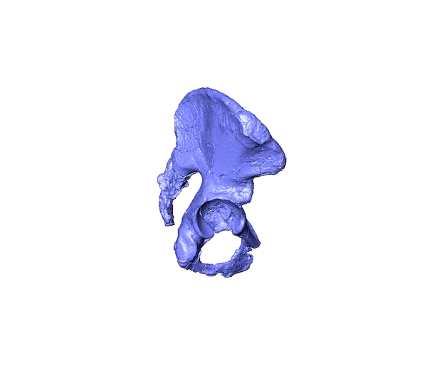
The paucity of well-preserved pelvises in the hominin fossil record has hindered robust analyses of shifts in critical biological processes throughout human evolution. The Kebara 2 pelvis remains one of the best preserved hominin pelvises, providing a rare opportunity to assess Neanderthal pelvic morphology and function. My co-authors and I created two new reconstructions of the Kebara 2 pelvis created from CT scans of the right hip bone and sacrum. First, we virtually reconstructed the right hip bone and the sacrum by repositioning the fragments of the hip bone and sacrum. Then, we created a mirrored copy of the right hip bone to act as the left hip bone. Next, we 3D printed the three bones and physically articulated them and finally, we used fiducial points collected from the physically articulated models to articulate the hip bones and sacrum in virtual space. Our objectives were to (1) reposition misaligned fragments, particularly the ischiopubic ramus; (2) create a 3D model of a complete pelvis; and (3) assess interobserver reconstruction variation. These new reconstructions show that, in comparison with previous measurements, Kebara 2 possessed a higher shape index (maximum anteroposterior length/maximum mediolateral width) for the pelvic inlet and perhaps the outlet and a more anteriorly positioned sacral promontory and pubic symphysis relative to the acetabula. The latter differences result in a lower ratio between the distances anterior and posterior to the anterior margins of the acetabula. Generally, the new reconstructions tend to accentuate features of the Kebara 2 pelvis - the long superior pubic ramus and anteriorly positioned pelvic inlet - that have already been discussed for Kebara 2 and other Neanderthals.
|
|
Adegboyega, M. T., Stamos, P. A., Hublin, J. J., & Weaver, T. D. (2021). Virtual reconstruction of the Kebara 2 Neanderthal pelvis. Journal of Human Evolution, 151, 102922. doi.org/10.1016/j.jhevol.2020.102922
Geometric morphometric analyses of shape variation in adult human pubic symphyses
|
|
The pubic symphysis is a secondary cartilaginous joint – a fibrocartilage and hyaline joint usually at the midline – that connects the left and right pubic bones to complete the anterior margin of the pelvic aperture. It is also responsible for absorbing shock during the transfer of the load from the upright trunk to the hips during locomotion and other weight bearing activities. Because cartilage is not preserved in fossil remains, our understanding of the morphology of the interpubic space is reliant on studies of modern humans.
I investigated the morphology of the pubic symphysis in adult modern humans from 103 adults by employing geometric morphometrics to assess shape variation across various anthropometric variables. I then trained 2-stage-least-squares linear regression models on these variables to predict pubic symphysis shape. To identify the strongest model, I compared the prediction errors generated through a process of leave one out cross validation. To visualize the performance of the best model, I created wireframe models of the predicted shapes from this model with its observed shape and I also created visualizations to show the effects of each predictor on the pubic symphysis shape. This study suggest that quantitatively predictive modeling could help to systematically estimate the shape of the pubic symphysis in humans. This method could potentially be applied along with other reconstruction techniques to improve fossil pelvic reconstructions by including an estimate for the shape of the bony and cartilaginous symphysis. |
Predicting the shape, position, and orientation of innominates in the pelvic girdle
|
I employed geometric morphometric techniques and analytical tools such as reduced rank regression modelling to accurately predicts the transformation indices that it would take to convert one hip bone to the shape, position and orientation of the other side. My model was tested on the modern human sample that I used in the previous study, and I collected new landmark data to suite the purposes of this study. I evaluated my model through leave one out modelling whereby a single individual was left out and the model was asked to predict what that individual’s left and right hip bones should look like, and how they should be positioned. My predictions of modern human hips within the training set were very impressive so I wanted to evaluate how well this same model would perform on fossil pelvises which are morphologically different to us. To achieve this, I introduced shape data for one hip bone from both of my Kebara reconstructions and used the model to predict the contralateral hipbone. When compared to the position of the hipbones in my reconstruction, I found the model to be accurate in its prediction.
|
|
